

 inglés
 francés
 español
 ruso
ruso coreano
 Japonés japonés
Exploring Innovation of Hyaluronic Acid Powder in Oral Care Formulations
Ácido hialurónico(HA), as a natural biopolymer, possesses excellent water-retaining capacity, lubricating properties ypermeation-regulating functions. It aids in maintaining tissue environment stability and supports healthy cellular metabolism. With ongoing application research, hyaluronic acid is demonstrating broad application prospects in the field deoral health and care.
Within oral care products, hyaluronic acid leverages its outstanding biocompatibility and unique physicochemical properties to serve as a premium ingredient across diverse categories including toothpaste, mouthwash, oral gels, and mucosal care solutions. It enhances product mildness, moisturising sensation, and overall user experience.
Primavera verde Technology is dedicated to supplying high-purity, multi-molecular-weight cosmetic-grade hyaluronic acid Polvo polvo polvo for the oral care sector. Our products boast exceptional stability and compatibility, meeting diverse development requirements for oral care formulations. This empowers brands to create safer, more comfortable, and functionally advanced oral health innovations.
1. Application Potential of Hyaluronic Acid Powder in OralHealth Care
As a natural polysaccharide, hyaluronic acid Polvo polvo polvo possesses excellent biocompatibility and mild physicochemical properties, making its application in oral care products increasingly noteworthy. It demonstrates significant potential in maintaining oral soft tissue health and improving the oral environment.
1.1 Complementing Daily Care, Enhancing the Health Experience
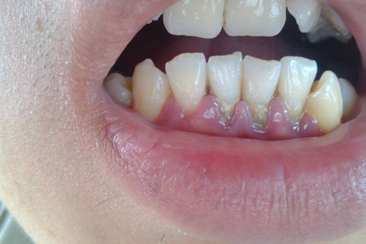
Research indicates hyaluronic acid can be incorporated into daily oral care routines to support gum health maintenance. Integrating hyaluronic acid-containing products with regular oral hygiene practices enhances the care experience and promotes overall oral environmental health [7]. From a tissue compatibility perspective, hyaluronic acid acts gently within the oral environment, making it suitable for daily care formulations.
1.2 Premium Ingredients Empowering OralCare Innovation
Owing to its unique molecular structure and physicochemical properties, hyaluronic acid powder serves as a superiorfunctional ingredient in toothpastes, mouthwashes, and oral gels, enhancing product usability and comfort. Suministros de tecnología de primavera verde high-purity cosmetic-grade hyaluronic acid raw materials, offering excellent stability and compatibility. These meetdiverse development needs for oral care products, enabling brands to create gentler, more effective oral health solutions.
2 Innovative Applications of Hyaluronic Acid in Oral Care
As a bioactive component naturally present in periodontal connective tissue,Polvo de ácido hialurónico possesses excellent biocompatibility and unique physicochemical properties. Its application in premium oral care products has garnered significant attention in recent years . Its superior moisturising properties, film-forming capabilities, and tissue affinity offer new avenues for innovative oral health product development.
Research indicates hyaluronic acid powder can serve as an adjunct ingredient in daily oral hygiene routines. When incorporated into conventional oral cleansing, products containing hyaluronic acid enhance the care experience and promote overall oral health. Xu Yi et al. [9] demonstrated that combining hyaluronic acid with professional cleaning methods delivers a more comfortable oral experience.
As a natural component of the extracellular matrix, hyaluronic acid regulates local hydration and maintains tissue moisture. Ácido hialurónico de alto peso molecular also forms protective films, contributing to a healthier oral environment. Multiple studies indicate that incorporating hyaluronic acid into oral care routines delivers a more comprehensive care experience.
Green Spring Technology specialises in supplying dental-grade hyaluronic acid powders. Our products feature high purity, multiple molecular weight options, and excellent compatibility, enabling broad application in toothpaste, mouthwash, oral gels, and other formulations. This empowers brand clients to develop more competitive and innovative oral care solutions.
3. Application Potential of Hyaluronic Acid in Implant Care
As a natural biopolymer, hyaluronic acid demonstrates broad application prospects in premium oral care—particularly peri-implant care—due to its exceptional biocompatibility and unique physicochemical properties. Its outstanding moisturising and film-forming capabilities provide a gentle care environment for the peri-implant region.
Research indicates that incorporating hyaluronic acid-containing products into routine oral hygiene enhances the overall care experience. Zhang Li et al.'s [11] study demonstrated that adding hyaluronic acid components yields positive care outcomes for the peri-implant region. Compared to mechanical cleaning alone, products incorporating hyaluronic acid exhibit superior comprehensive advantages in improving the local environment.
Within professional oral care, researchers have explored combining hyaluronic acid powder with other care ingredients. Experiments indicate that hyaluronic acid-based care regimens offer users a more comfortable experience, delivering comparable efficacy to certain traditional care ingredients while being gentler.
4 Applications and Prospects of Hyaluronic Acid in Post-Oral Surgical Care
Ácido hialurónico powder demonstrates significant application potential in post-oral surgical care due to its excellent biocompatibility and moisturising properties. Research indicates it provides positive microenvironmental support during post-extraction care, promoting orderly tissue recovery.
Experiments demonstrate that application of high molecular weight hyaluronic acid gel results in more orderly local tissue arrangement, suggesting it may create favourable conditions for wound healing. Further studies confirm that hyaluronic acid can regulate the local microenvironment to provide nutritional support for tissues, aiding the healthy recovery process.
Green Spring Technology supplies dental-grade hyaluronic acid powders characterised by high purity, multiple molecular weight options, and excellent stability. These are suitable for developing products such as oral gels and post-operative care solutions, providing reliable support for brand clients innovating premium oral care offerings.
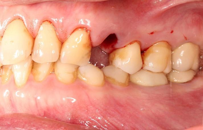
5 Applications of Hyaluronic Acid in Implant Care
Hyaluronic acid powder demonstrates application potential in post-operative care for dental implants due to its superior moisturising and film-forming properties. Its excellent biocompatibility aids in creating a suitable microenvironment, offering innovative directions for post-operative care products.
Research indicates that hyaluronic acid products can support wound condition maintenance in the early post-operative phase. By forming a protective film, they maintain a moist environment and enhance comfort. Although study results vary due to factors such as evaluation methods, its value as a functional raw material remains noteworthy.
6 Innovative Applications of Hyaluronic Acid Powder in Oral Mucosal Care
Hyaluronic acid powder, with its exceptional moisturising and film-forming properties, has become a key functional ingredient in oral mucosal care products. Its excellent biocompatibility delivers a gentle yet effective care experience for the oral cavity.
Research indicates that oral care products containing hyaluronic acid powder significantly enhance user comfort. Experiments demonstrate that after using products with 0.2% hyaluronic acid, users' oral conditions show marked improvement. Hyaluronic acid forms a breathable protective film on mucosal surfaces, effectively shielding against external irritants while maintaining localised moisture.
Green Spring Technology leverages advanced bio-extraction and molecular modification techniques to develop a series of hyaluronic acid powder Soluciones solucionesspecifically for oral care. Our products utilise precise molecular weight control technology to fulfil diverse functional requirements—from low molecular weight rapid penetration to high molecular weight long-lasting film-forming properties—offering formulators flexible and versatile options.

This hyaluronic acid powder exhibits exceptional sensory properties, significantly enhancing the smoothness and skin-adherence of end products. It demonstrates excellent formulation compatibility and system stability, suitable for diverse formulations including mouthwashes, oral gels, and mucosal care solutions. All products adhere to internationally recognised quality control standards, undergoing rigorous quality testing and safety assessments to ensure batch-to-batch consistency and regulatory compliance.
Please contact us at helen@greenspringbio.com or WhatsApp: +86 13649243917 for product specifications and quotations. Green Spring Technology provides specialised, bespoke raw material solutions to advance the technological evolution and market innovation of oral care products.
referencias
[1] Huang Sili, Guo Xueping, Yang Guilan, et al. Recientes avances en la aplicación del ácido hialurónico [J]. Food and Medicine, 2009,11(1): 50-53.
[2] Largo largo X, Chen G, Cheng AH, et Al. Un ensayo aleatorizado con- trolled trial of superior and Inferior inferior Articulación temporomandibular Espacio espacio Inyección de inyección Con ácido hialurónico en el tratamiento de la anterior Disco disco El desplazamiento sin Reducción [J]. J Los demás Surg, 2009, 67(2):357-361.
[3] Escoda-Francoli J, Vzquez-Delgado E, Gay-Escoda C.Scientific evidence on the usefulness of intraarticular hyaluronic acid injection in the management of temporo- mandibular Disfunción [J]. Med Oral Patol Oral Cir Bucal, 2010, 15(4):e644-e648.
[4] Li Chunjie, Zhang Yifan, Jia Yuanyuan, et al. Una revisión sistemática de un ensayo clínico controlado aleatorizado sobre el tratamiento de la disfunción de la articulación temporomandibular con hialuronato de sodio [J]. West China Journal of Stomatology, 2011, 29(5): 488-493.
[5] Liu Peicai, Wang Dong, Peng Cheng, et al. Efectos del hialuronato de sodio sobre las metaloproteinasas de la matriz 2 y 3 en el líquido sinovial de pacientes con osteoartritis de la articulación temporomandibular [J]. Chinese Journal of Stomatological Research: Electronic Edition, 2011, 5(4): 356-360.
[6] Zhao Jigang, Peng Guoguang, Liang Jingzhang, et al. Estudio clínico sobre la prevención de complicaciones postoperatorias de fractura condilar por lavado articular e inyección de hialuronato de sodio [J]. Journal of Modern Stomatology, 2011, 25(2): 105-107.
[7] Wu Yafei, Huang Jiao, Xu Yi, et al. El papel del gel de Gengigel en el tratamiento de la gingivitis inducida por placas [J]. Journal of Practical Stomatology, 2005, 21(4): 540-542.
[8] Sukumar S, Drizhal I. ácido hialurónico y periodontitis [J]. Acta Medica (Hradec Králové), 2007, 50(4): 225-228.
[9] Xu Yi, Frentzen M, Jerv → e-Storm P-M. El papel del ácido hialurónico en el tratamiento de la periodontitis crónica [J]. West China Journal of Stomatology, 2004, 22(1): 32-34.
-
anterior
Exploring Innovation with Hyaluronic Acid Powder in Skincare Products
-
siguiente
Hyaluronic Acid Elevates Innovation in Oral Care Products